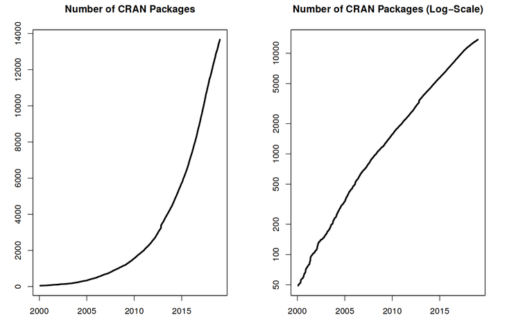
画像1/1: ‘Two graphs showing the exponential increase of the number of CRAN packages from 2000 to 2018. The figure on the right shows linear trend for the log of number of packages.’
Exponential increase of the number of packages available on CRAN, the Comprehensive R Archive
Network. From the R Journal, Volume 10/2, December 2018.
図の2
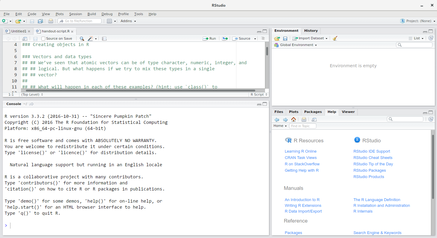
画像1/1: ‘Screenshot of the RStudio interface showing the typical four panel setup.’
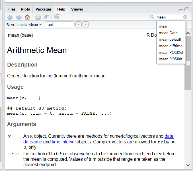
RStudio interface screenshot. Clockwise from top left: Source,
Environment/History, Files/Plots/Packages/Help/Viewer, Console.
図の3
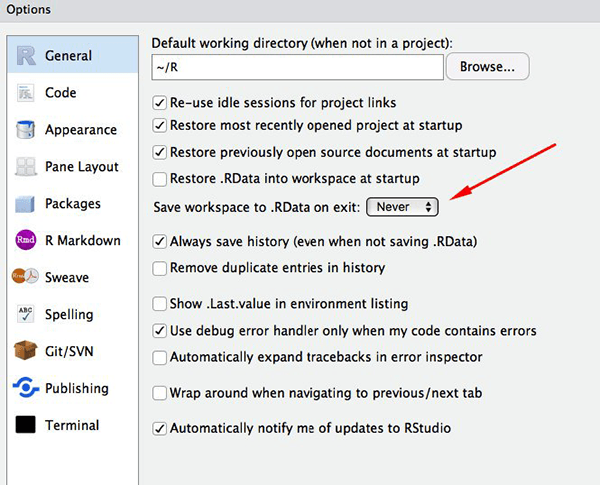
画像1/1: ‘Screenshot of the RStudio General Options panel.’
Set ‘Save workspace to .RData on exit’ to ‘Never’
図の4
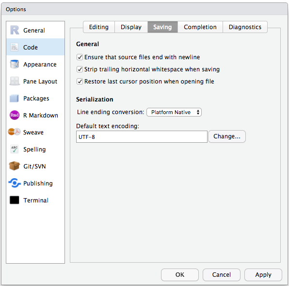
画像1/1: ‘Screenshot of the RStudio Code Options panel.’
Set the default text encoding to UTF-8 to save us headache in the coming
future. (Figure from the link above).
図の5
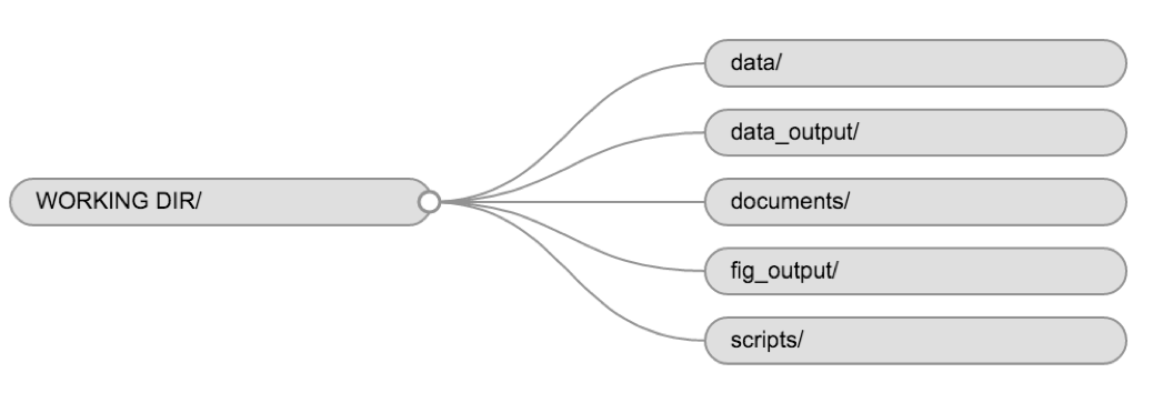
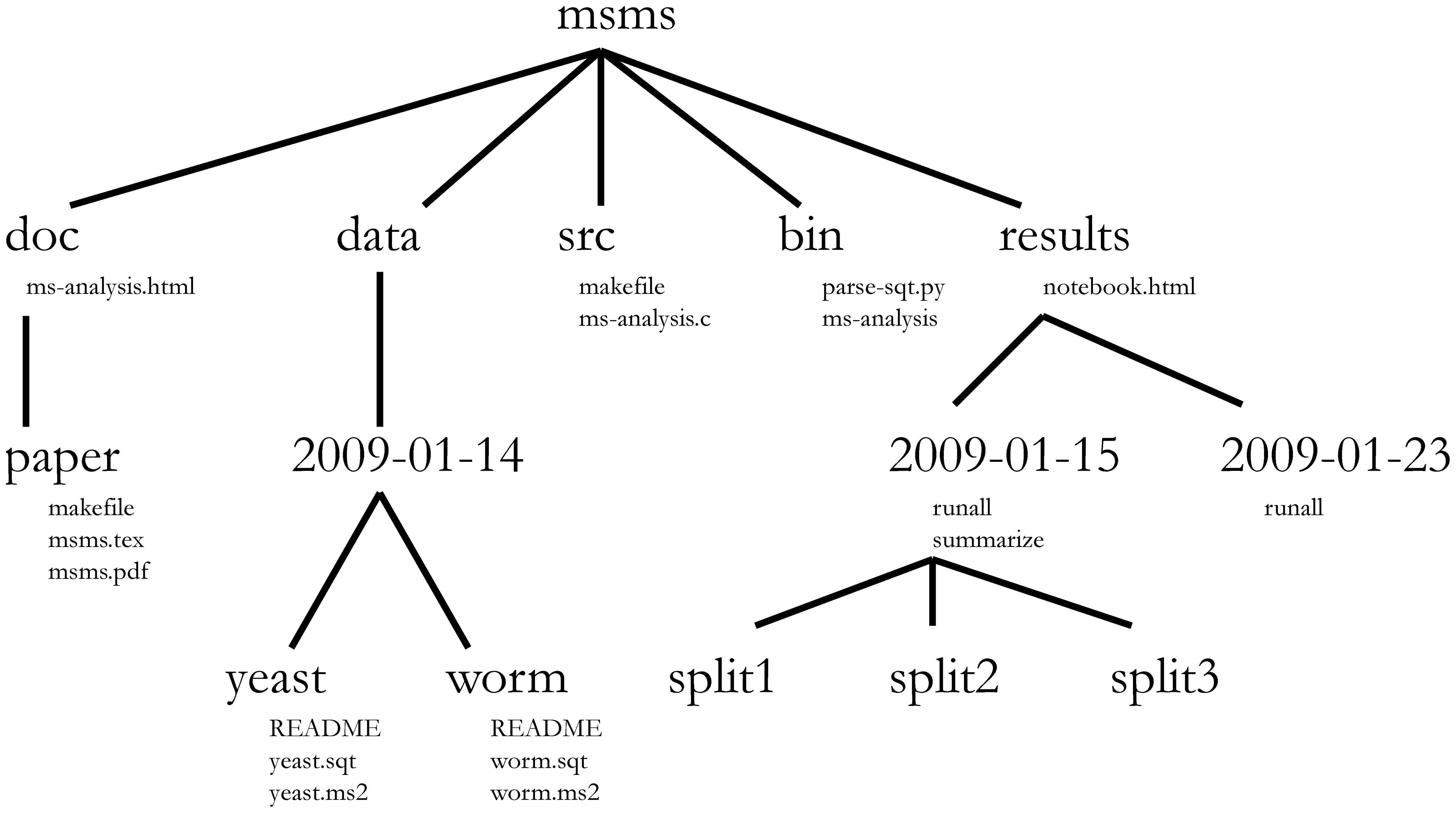
画像1/1: ‘Flow diagram showing the structure of the working directory and various directory of a typical RStuio project.’
Example of a working directory structure.
図の6
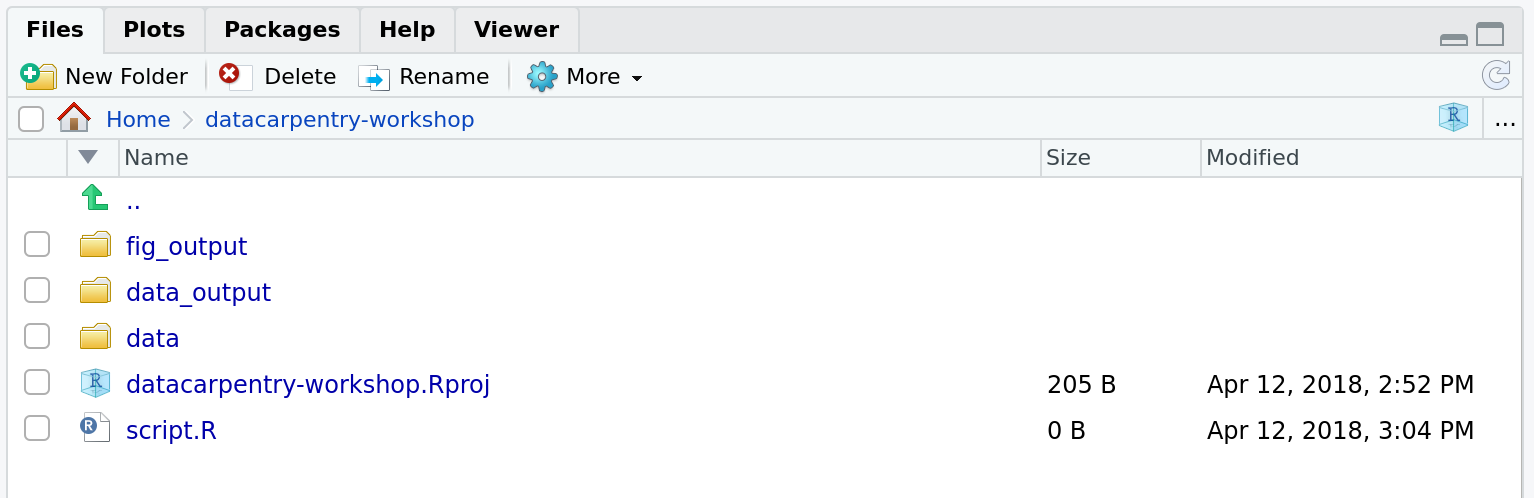
画像1/1: ‘Screenshot of RStudio's Files panel.’
How it should look like at the beginning of this lesson
図の7
画像1/1: ‘Figure form Nobel et al. (2009) showing a complex bioinformatics project structure.’
Directory structure for a sample bioinformatics project.
図の8
画像1/1: ‘Fake book cover showing a kitten with title 'Changing Stuff and Seeing What Happens.'’
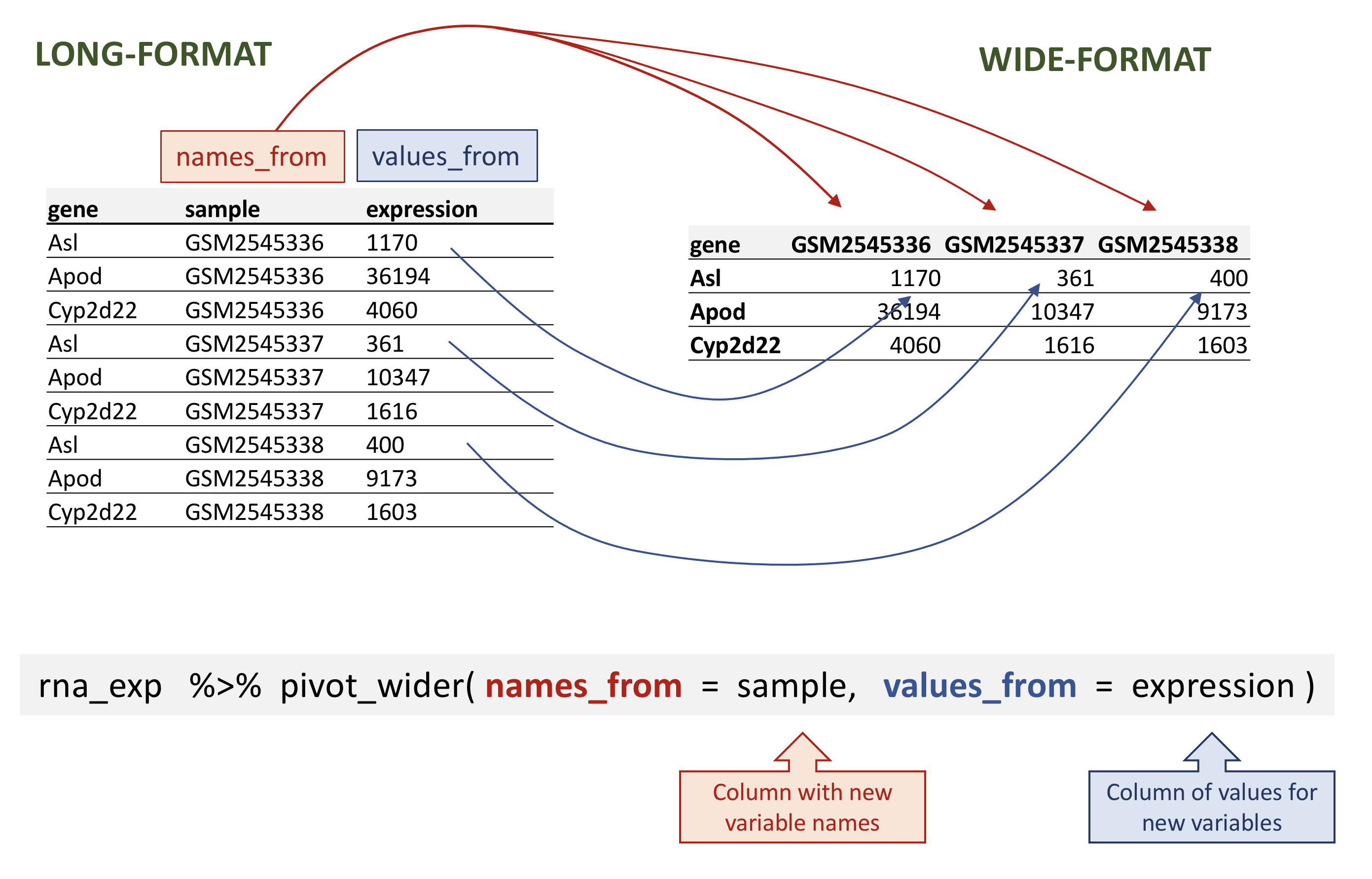
画像1/1: ‘Figure showing the long table format on the left and wide table format on the right and arrows illustrating how the values in the 'sample' column on the left have become column names on the right and how the values in the 'expression' column on the left have become values on the the right. Below is the call to 'pivot_wider()' with annotations pointing to the 'sample' and 'expression' function arguments.’
Wide pivot of the rna data.
図の2
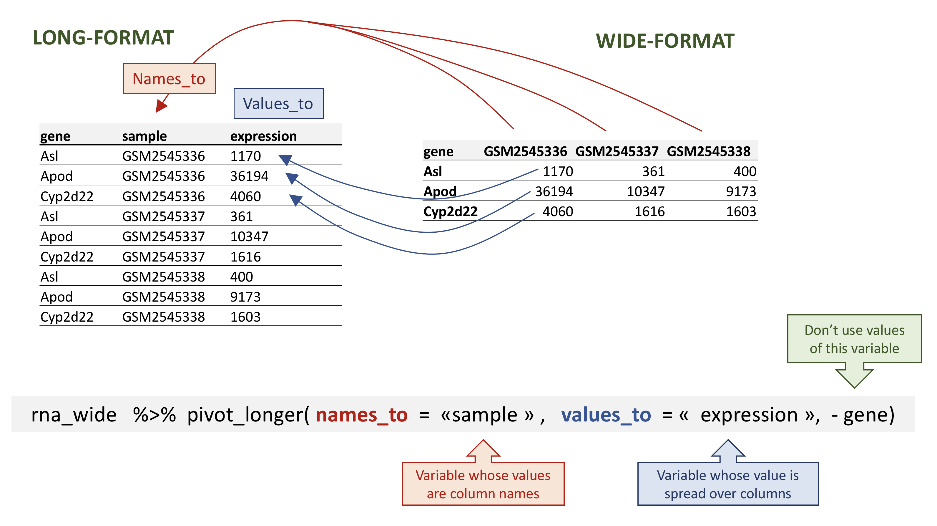
画像1/1: ‘Figure showing the long table format on the left and wide table format on the right and arrows illustrating how the column names on the left have become a new column 'sample' on the left and the values in the wide table on the right have become a new column 'expression' on the left. Below is the call to 'pivot_wider()' with annotations pointing to the 'sample', 'expression' and the '-gene' arguments.’
Long pivot of the rna data.
図の3
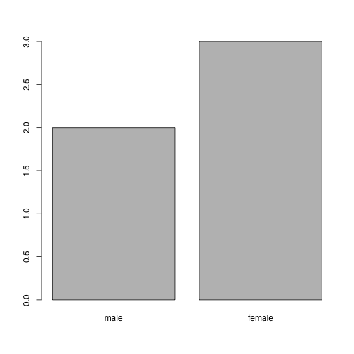
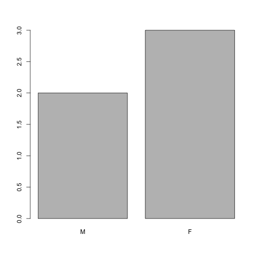
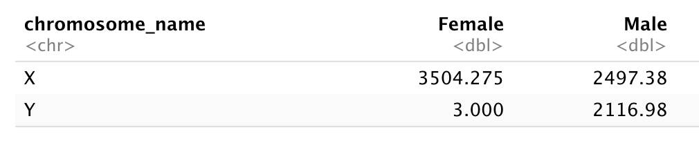
画像1/1: ‘A two by two table with X and Y row names and Female and Male column names showing the total counts for each X/Y and Female/Male combination. The Y/Female combinations shows 3 counts, while all other counts are above 2000 counts.’
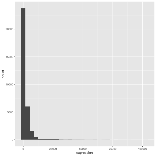
画像1/1: ‘Default histogram produced by ggplot() and geom_histogram() for the expression data.’
図の2
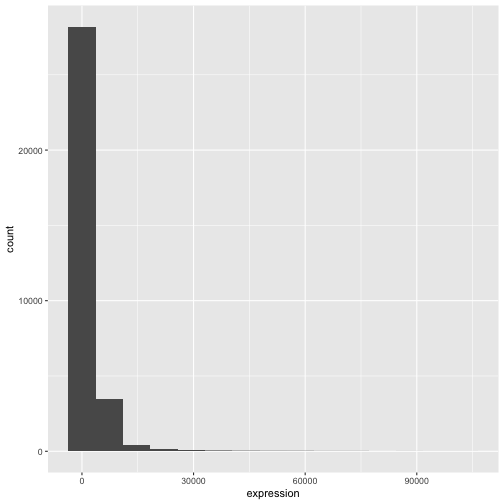
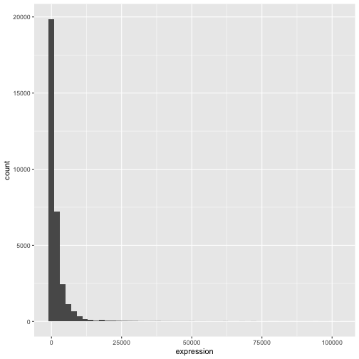
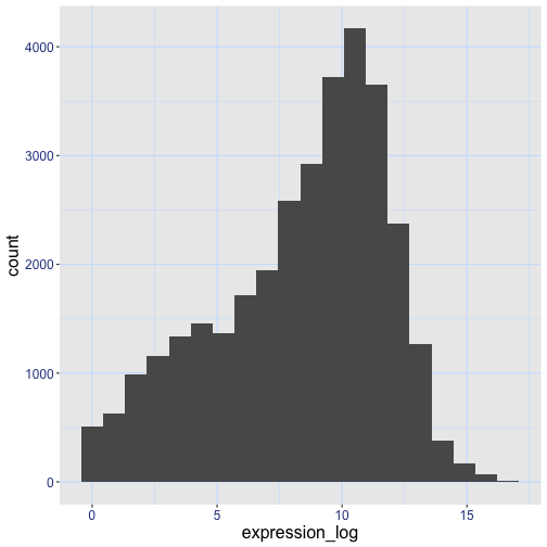
画像1/1: ‘Histograms produced by ggplot() and geom_histogram() for the expression data with bin set of 15 (top) and binwith set to 2000 (bottom).’
図の3
画像1/1: ‘Histograms produced by ggplot() and geom_histogram() for the expression data with bin set of 15 (top) and binwith set to 2000 (bottom).’
図の4
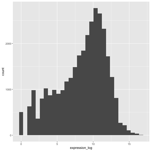
画像1/1: ‘Histogram produced by ggplot() and geom_histogram() for the pre-computed log of expression.’
図の5
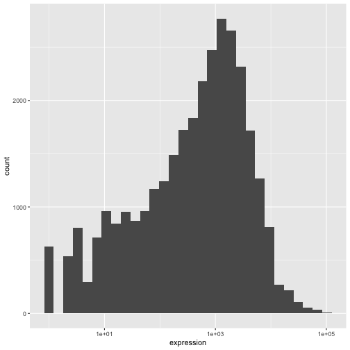
画像1/1: ‘Histogram produced by ggplot(), geom_histogram() and scale_x_log10() for the log of expression.’
図の6
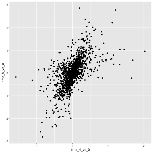
画像1/1: ‘Scatter plot produced by ggplot() and geom_point() comparing the log-foldchanges computed above. All dots are black.’
図の7
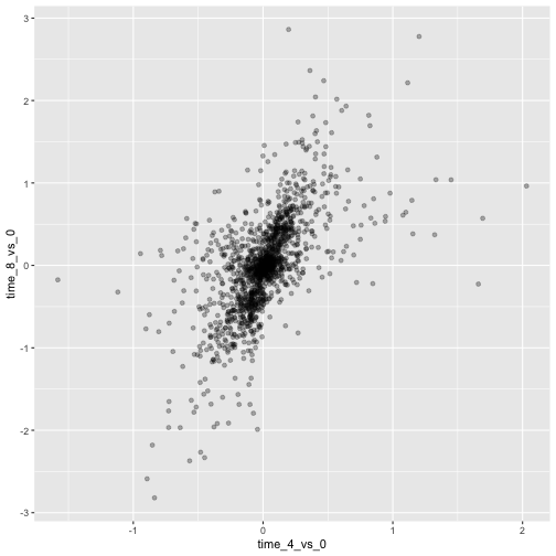
画像1/1: ‘Scatter plot produced by ggplot() and geom_point() comparing the log-foldchanges computed above. All dots are semi-transparent black.’
図の8
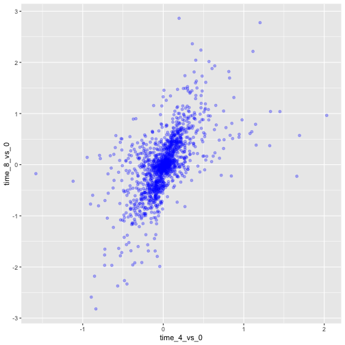
画像1/1: ‘Scatter plot produced by ggplot() and geom_point() comparing the log-foldchanges computed above. All dots are semi-transparent blue.’
図の9
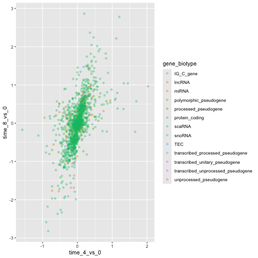
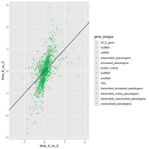
画像1/1: ‘Scatter plot produced by ggplot() and geom_point() comparing the log-foldchanges computed above. Dots are colour-coded based on the gene's biotype.’
図の10
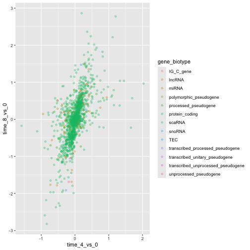
画像1/1: ‘Scatter plot produced by ggplot() and geom_point() comparing the log-foldchanges computed above. Dots are colour-coded based on the gene's biotype.’
図の11
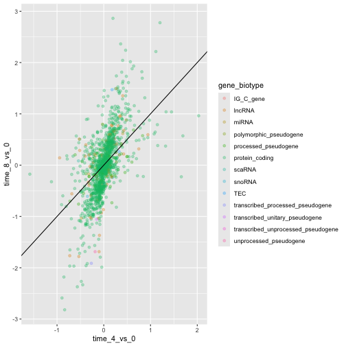
画像1/1: ‘Scatter plot produced by ggplot() and geom_point() comparing the log-foldchanges computed above. Dots are colour-coded based on the gene's biotype. A black line of slope 1 crossing the origin was added by geom_abline().’
図の12
画像1/1: ‘Scatter plot produced by ggplot() and geom_point() comparing the log-foldchanges computed above. Dots are colour-coded based on the gene's biotype. A black line of slope 1 crossing the origin was added by geom_abline().’
図の13
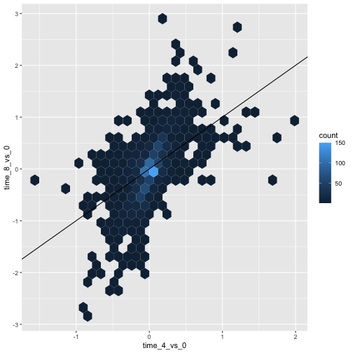
画像1/1: ‘Scatter plot produced by ggplot() and geom_hexbin() comparing the log-foldchanges computed above shows hexagons coloured based on the underlying dot density. A black line of slope 1 crossing the origin was added by geom_abline().’
図の14
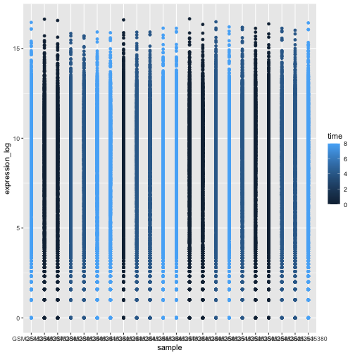
画像1/1: ‘Figures showing a stretch of overlapping points indicating the log of expression + 1 for each sample. The points are coloured with different shades of blue for samples collected at different time points.’
図の15
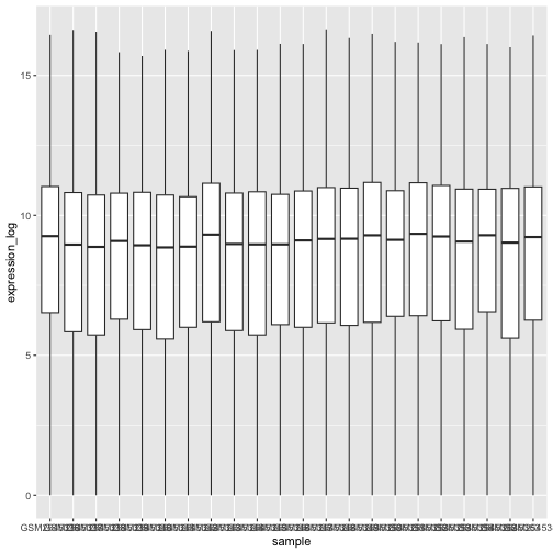
画像1/1: ‘Boxplot showing the distribution of log expression + 1 values for each sample, as produced by geom_boxlpot(). Each boxplot is filled with white colour.’
図の16
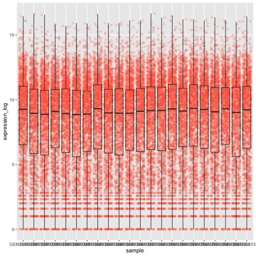
画像1/1: ‘Boxplot and dots showing the distribution of log expression + 1 values for each sample, as produced by geom_boxlpot(). Each boxplot is transparent and the jittered dots are semi-transparent tomato-coloured and behind the boxpots.’
図の17
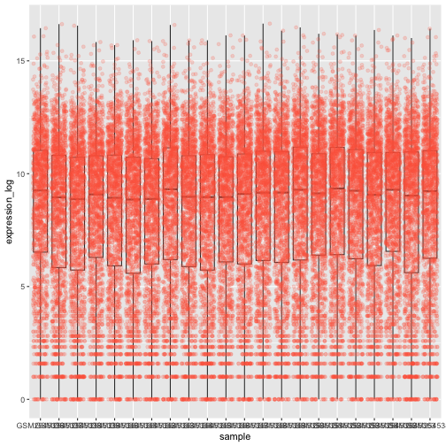
画像1/1: ‘Boxplot and dots showing the distribution of log expression + 1 values for each sample, as produced by geom_boxlpot(). Each boxplot is transparent and the jittered dots are semi-transparent tomato-coloured. This time, the boxplots are behind the dots.’
図の18
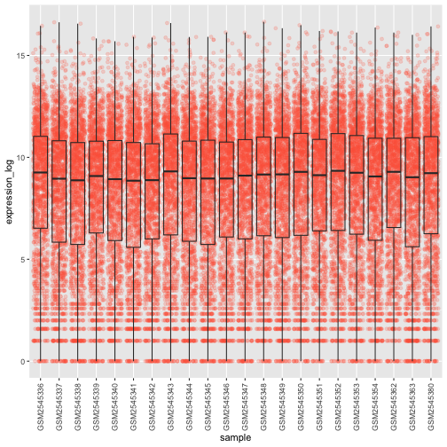
画像1/1: ‘Boxplot and dots showing the distribution of log expression + 1 values for each sample, as produced by geom_boxlpot(). Each boxplot is transparent and the jittered dots are semi-transparent tomato-coloured. The sample labels are displayed vertically and readable.’
図の19
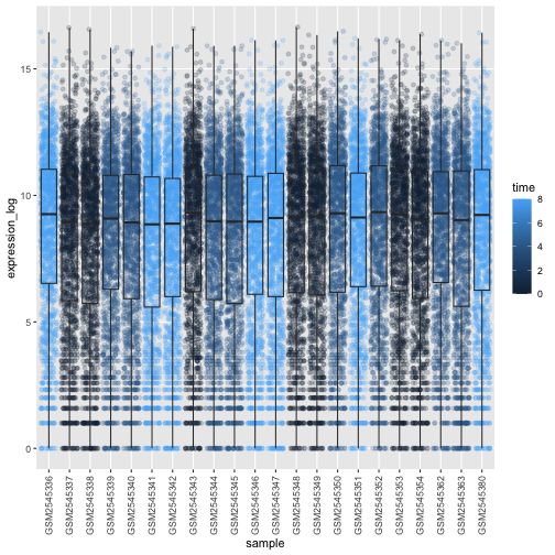
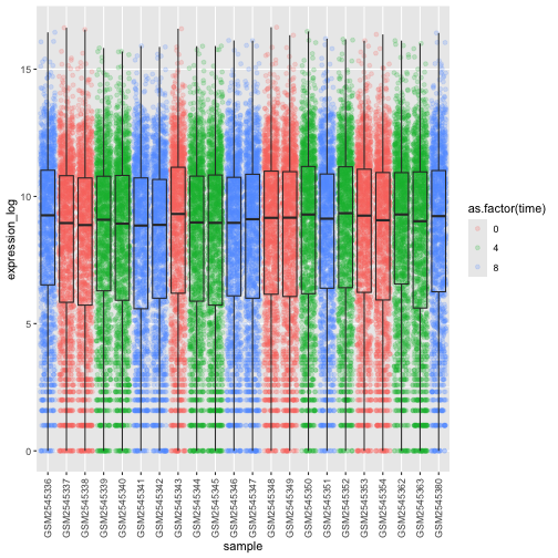
画像1/1: ‘Boxplot and dots showing the distribution of log expression + 1 values for each sample, as produced by geom_boxlpot(). On the first figure, each boxplot is transparent and the jittered dots are semi-transparent and coloured in different shares of blue. On the second figures, each boxplot is transparent and the jittered dots are semi-transparent and coloured red, green and blue.’
図の20
画像1/1: ‘Boxplot and dots showing the distribution of log expression + 1 values for each sample, as produced by geom_boxlpot(). On the first figure, each boxplot is transparent and the jittered dots are semi-transparent and coloured in different shares of blue. On the second figures, each boxplot is transparent and the jittered dots are semi-transparent and coloured red, green and blue.’
図の21
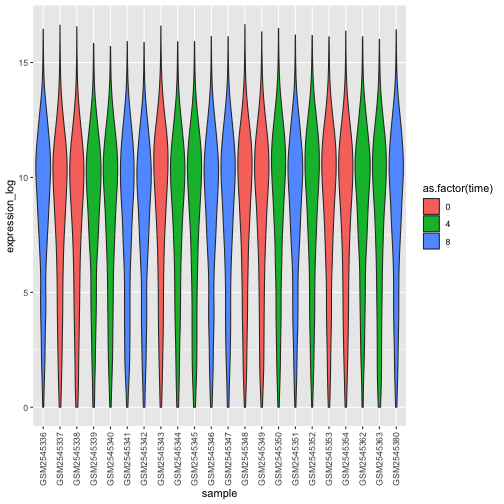
画像1/1: ‘Violin plot showing the distribution of log expression + 1 values for each sample, as produced by geom_violin(). Each boxplot is transparent and the jittered dots are semi-transparent and coloured red, green and blue.’
図の22
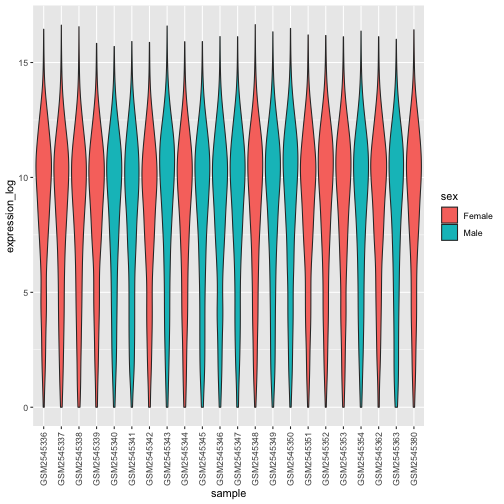
画像1/1: ‘Violin plot showing the distribution of log expression + 1 values for each sample, as produced by geom_violon(). Each violin plot is coloured in red or blue depending on the sex variable.’
図の23
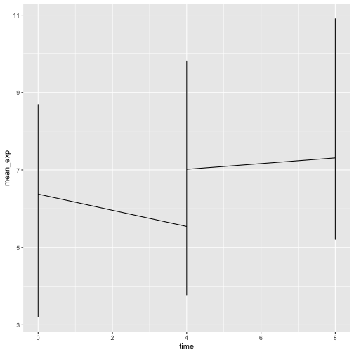
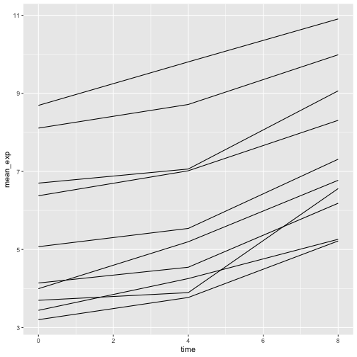
画像1/1: ‘Line plot, as produced by geom_line(), but the lines are clearly not show what we expect.’
図の24
画像1/1: ‘Line plot, as produced by geom_line(), with 10 lines showing increasing expression values over time.’
図の25
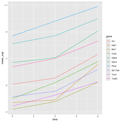
画像1/1: ‘Line plot, as produced by geom_line(), with 10 colour-coded lines showing increasing expression values over time.’
図の26
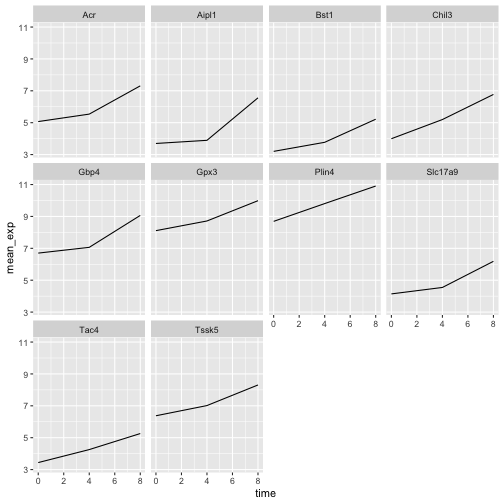
画像1/1: ‘Line plot, as produced by geom_line(), with 10 sub-plots/facets, each showing one line with increasing expression values over time. All y-scales are identical.’
図の27
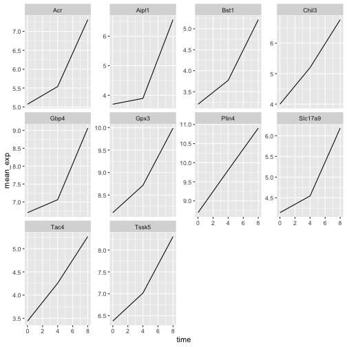
画像1/1: ‘Line plot, as produced by geom_line(), with 10 sub-plots/facets, each showing one line with increasing expression values over time. y-scales are now adapted to the expression range for each facet/gene.’
図の28
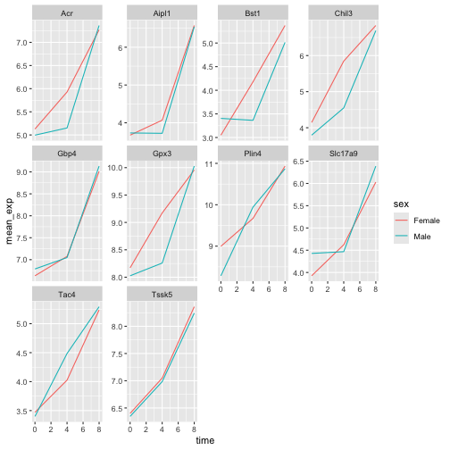
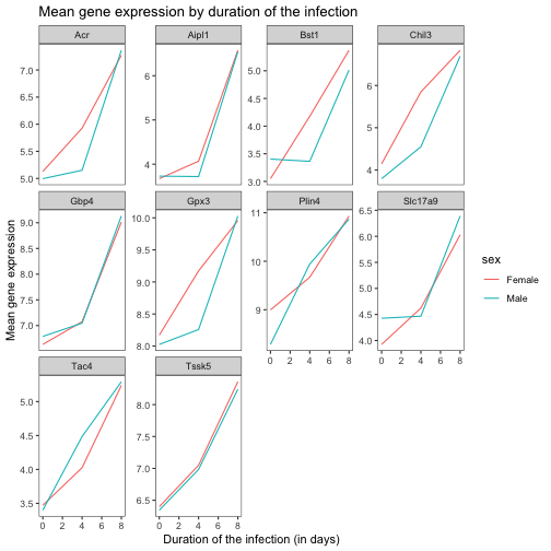
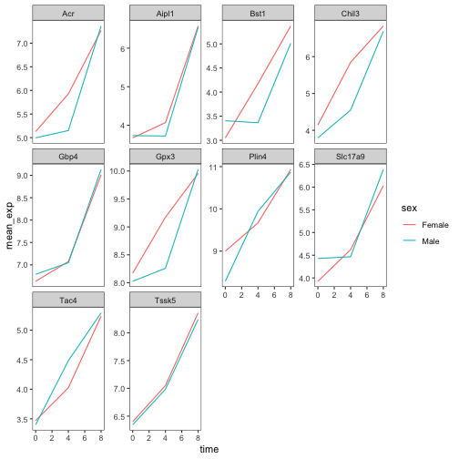
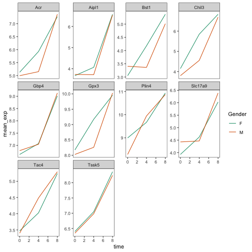
画像1/1: ‘Line plot, as produced by geom_line(), with 10 sub-plots/facets, each showing two coloured lines (red for Female and blue for Make) with increasing expression values over time.’
図の29
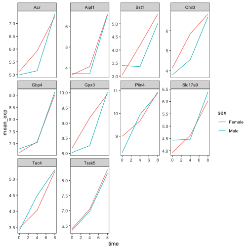
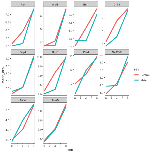
画像1/1: ‘Line plot, as produced by geom_line(), with 10 sub-plots/facets, each showing two coloured lines (red for Female and blue for Make) with increasing expression values over time. The figure background is now white.’
図の30
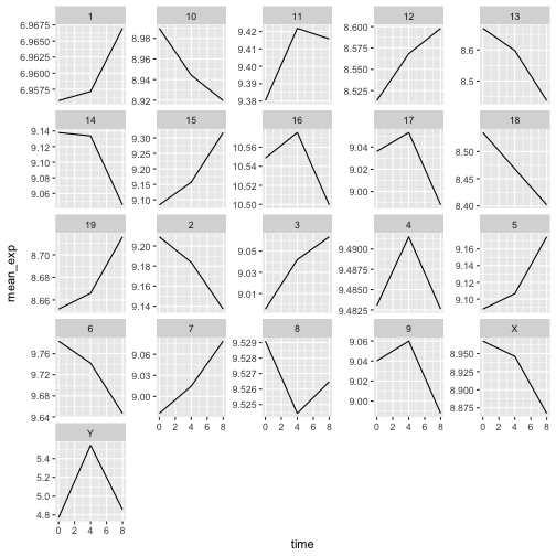
画像1/1: ‘Line plot, as produced by geom_line(), with 21 sub-plots/facets, each showing one line with expression values over time for each chromosome.’
図の31
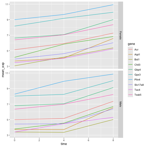
画像1/1: ‘Two line plots above each other, each showing 10 lines coloured by gene. The top facet shows the expression values for Female sample, the bottom one for Male samples.’
図の32
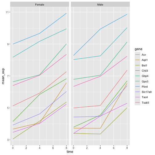
画像1/1: ‘Two line plots next to each other, each showing 10 lines coloured by gene. The left facet shows the expression values for Female sample, the right one for Male samples.’
図の33
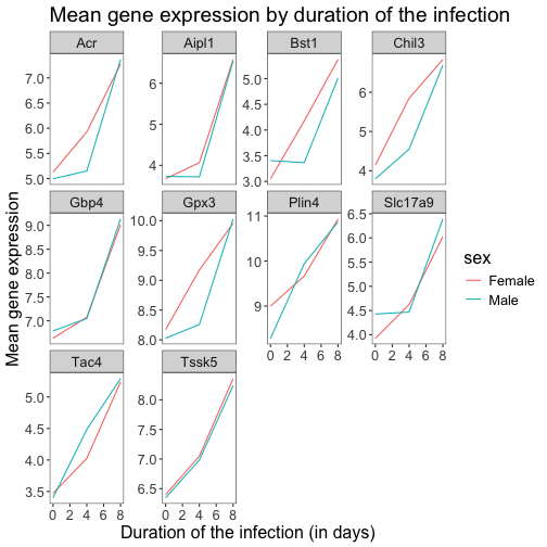
画像1/1: ‘Line plot with white background and custom title and axis legends.’
図の34
画像1/1: ‘Line plot with white background and larger custom title and axis legends.’
図の35
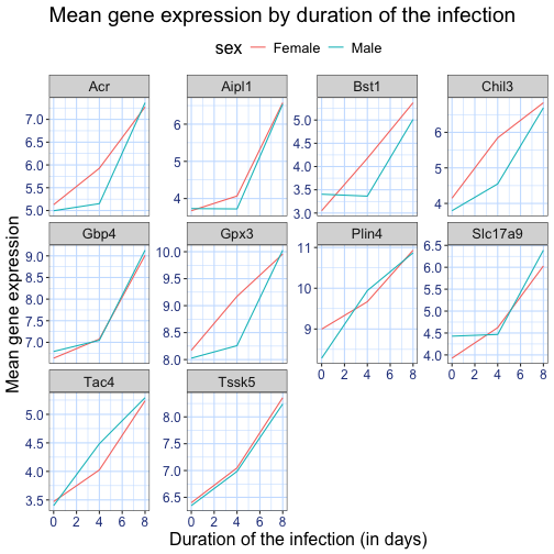
画像1/1: ‘Line plot with white background and larger custom title and axis legends and blue grid.’
図の36
画像1/1: ‘Custrom 'blue_theme() showing a blue grid and blue axis legends.’
図の37
画像1/1: ‘Plain facetted line plot, as produced by theme_bw() and a blank grid above.’
図の38
画像1/1: ‘Plain facetted line plot, as produced by theme_bw() and a blank grid with wide lines.’
図の39
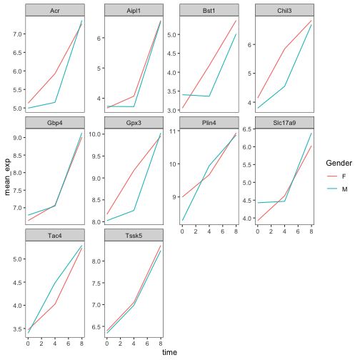
画像1/1: ‘Plain facetted line plot, as produced by theme_bw() and a blank grid with renamed colour labels.’
図の40
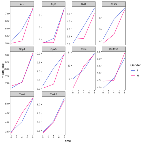
画像1/1: ‘Plain facetted line plot, as produced by theme_bw() and a blank grid with a different color palette and renamed colour labels.’
図の41
画像1/1: ‘Plain facetted line plot, as produced by theme_bw() and a blank grid with a manually-set colors and renamed colour labels..’
図の42
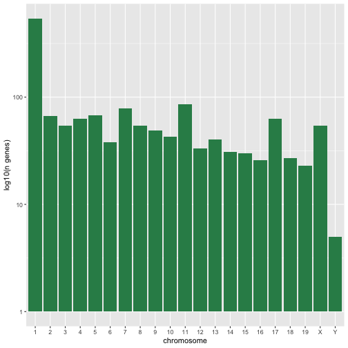
画像1/1: ‘A simple histogram with greed bars showing the log10 number of genes per chromosome.’
図の43
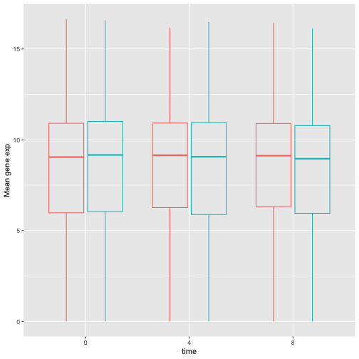
画像1/1: ‘Transparently-filled red and blue boxplots for Female and Male expression values for different time points.’
図の44
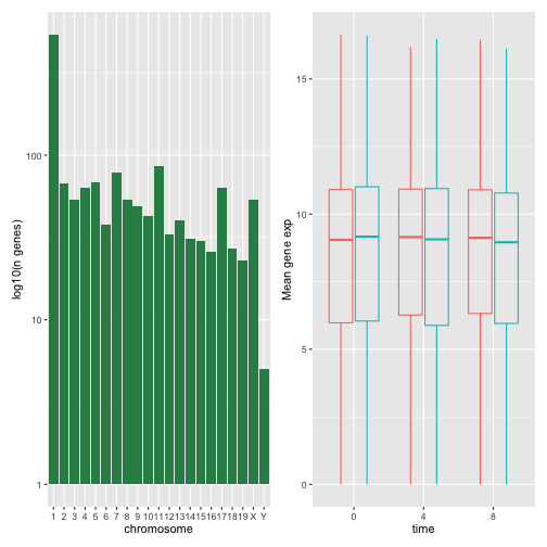
画像1/1: ‘A composed figure show the histogram (left) and boxplots (right) next to each other.’
図の45
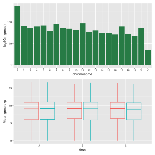
画像1/1: ‘A composed figure show the histogram (top) and boxplots (bottom) on top or each other.’
図の46
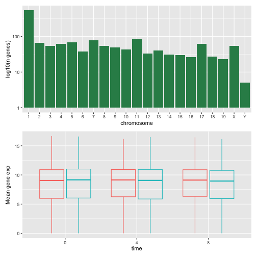
画像1/1: ‘A composed figure show the histogram (top) and boxplots (bottom) on top or each other.’
図の47
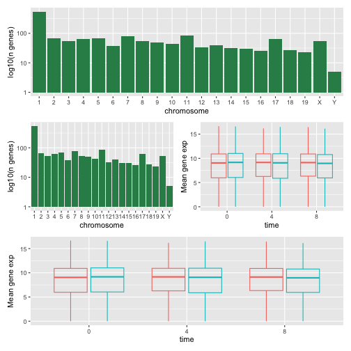
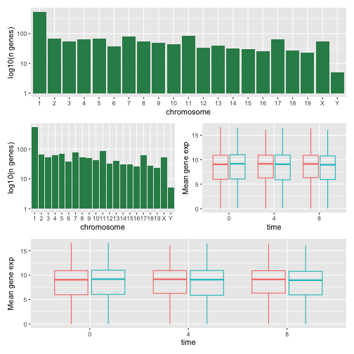
画像1/1: ‘A figure composed of 4 plots with on top the histogram, in the middle the histogram and the boxpot, side-by-side, and at the bottom, the boxplot.’
図の48
画像1/1: ‘As above, a figure composed of 4 plots with on top the histogram, in the middle the histogram and the boxpot, side-by-side, and at the bottom, the boxplot.’
図の49
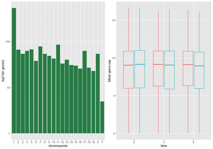
画像1/1: ‘A composed figure with the histogram (left) and boxplot (right) side-by-side.’
図の50
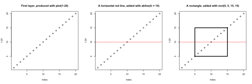
画像1/1: ‘The 'base' figures, side-by-side, showing (from left to right) 20 empty dots along the diagonal, same with a vertical red line, and same as the second one with a rectangle overlaid in the middle of the plot.’
Successive layers added on top of each other.
図の51
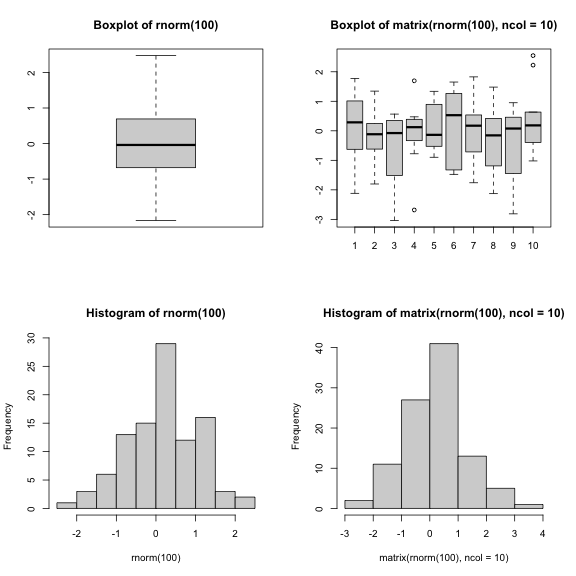
画像1/1: ‘A two-by-two composition of 2 boxplots at the top, and 2 histograms at the bottom.’
Plotting boxplots (top) and histograms (bottom) vectors (left) or a
matrices (right).
画像1/1: ‘Schematic representation of the SummarizedExperiment class illustrating the following slots: fowData and rowRanges, one assay or multiple assays, colData and metadata.’
図の2
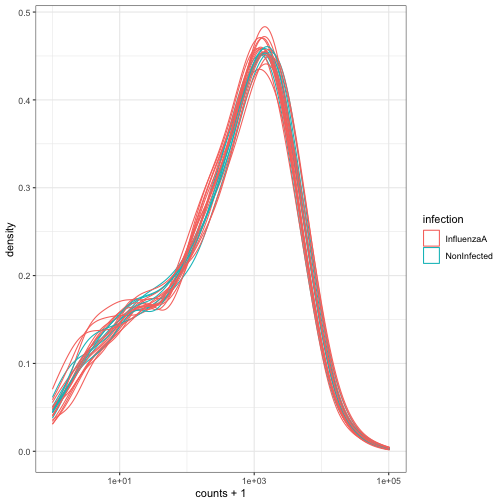
画像1/1: ‘Density plot showing log of expression counts + 1 density lines, one per samples, coloured based on the infection status.’